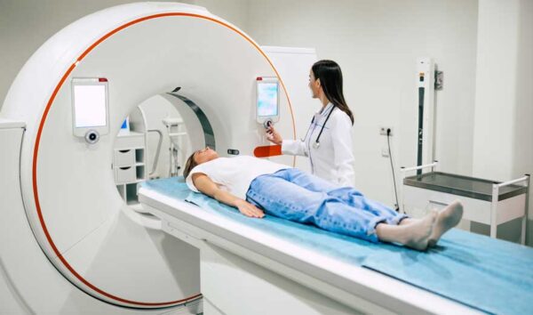
Radiology

Radiology, also known as diagnostic imaging, is a series of tests that take pictures or images of parts of the body. The field encompasses two areas — diagnostic radiology and interventional radiology — that both use radiant energy to diagnose and treat diseases.
Radiology is used for a wide range of conditions and is classified depending on the type of radiology and the exact imaging test used. The various imaging exams include:
- Radiographs: X-rays to look at bones, the chest, or the abdomen.
CT (Computed Tomography): A CT captures multiple x-ray angles of the patient using a doughnut-shaped machine, then creates computer-processed images. - MRI (Magnetic Resonance Imaging): An MRI uses magnetic fields and radio waves with computer processing to create images.
- Mammograms: Especially powered x-rays that look at breast tissues.
- Ultrasound: An ultrasound uses sound waves to create moving images that display on a monitor, commonly used for echocardiograms and examining the womb during pregnancy.
- Fluoroscopy: X-rays that make moving images of the body in real-time. This imaging is crucial for many procedures, especially those involving the gastrointestinal tract.
- Nuclear medicine: These are short-acting radioactive substances that generate light from bodily processes. A camera collects the light, so a computer can process it and develop an image.
Diagnostic radiology uses these imaging results to identify a wide range of problems, from broken bones to heart conditions and blood clots. Interventional radiology also uses imaging such as CT scans, MRI, and ultrasounds to guide medical procedures. Patients are typically awake during these procedures, whether it’s treating cancer, back pain, or liver and kidney problems. In some cases, interventional radiology eliminates the need for surgery and scopes.
